Home
/ Shoulder Muscles Diagram Labeled : Muscle Diagram Anatomy System Human Body Anatomy Diagram And Chart Images - The fascia merges with the periosteum (outer bone layer) of the humerus.
Shoulder Muscles Diagram Labeled : Muscle Diagram Anatomy System Human Body Anatomy Diagram And Chart Images - The fascia merges with the periosteum (outer bone layer) of the humerus.
Shoulder Muscles Diagram Labeled : Muscle Diagram Anatomy System Human Body Anatomy Diagram And Chart Images - The fascia merges with the periosteum (outer bone layer) of the humerus.. Shoulder anatomy includes the deltoid muscle, supraspinatus, infraspinatus and subscapularis. Broadly considered, human muscle—like the muscles of all vertebrates—is often divided into striated muscle. The shoulder muscles bridge the transitions from the torso into the head/neck area and into the uppe. Shoulder muscles diagram labeled : Related posts of shoulder muscles and tendons diagram muscle anatomy coloring book.
Shoulder muscles diagrams | 101 diagrams from www.101diagrams.com These muscles form the outer shape of the shoulder and underarm. Identify the muscle labeled as 1 in the diagram above: The shoulder has about eight muscles that attach to the scapula, humerus, and clavicle. The rotator cuff is a collection of muscles and tendons that surround the shoulder, giving it support and allowing a wide range of motion.
Shoulder Anatomy Labeled Hd Stock Images Shutterstock from image.shutterstock.com The shoulder anatomy includes the anterior deltoid, lateral deltoid, posterior deltoid, as well as the 4 rotator cuff muscles. Related posts of shoulder muscles labelled diagram muscles labeled front and back. The first muscle diagram labeled 2019 above gives you an illustration of the anatomy of the arm muscle. Assessment of the flexibility of certain muscles as the disease progresses, night pain becomes more common. Posted on april 26, 2016 by admin. Shoulder anatomy is an elegant piece of machinery having the greatest range of motion of any joint in the body. This flexibility is also what makes the shoulder prone to instability and injury. The fascia merges with the periosteum (outer bone layer) of the humerus.
Learn about these muscles, their origin and insertion points, and their functional anatomy.
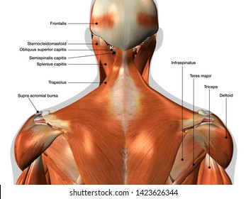
The rotator cuff is a collection of muscles and tendons that surround the shoulder, giving it support and allowing a wide range of motion. Subscapularis, supraspinatus, infraspinatus and teres minor. Anatomy organs human body anatomy anatomy and physiology anatomy male arm anatomy anatomy drawing shoulder muscle anatomy shoulder blade muscles arm muscle anatomy. The muscles of the shoulder are associated with movements at the shoulder joint. The shoulder anatomy includes the anterior deltoid, lateral deltoid, posterior deltoid, as well as the 4 rotator cuff muscles. Plus, exercises for training them. Explore and learn the muscles of the shoulder with our 3d interactive anatomy muscle atlas. Shoulder flexion is movement of the shoulder in a forward motion. Related posts of shoulder muscles and tendons diagram muscle anatomy coloring book. Shoulder muscles diagram labeled : The list of muscles and their functions are presented below. This diagram depicts muscle labeled diagram. Muscles of the shoulder :
Shoulder muscles move the shoulder blades and upper arm bones. The rotator cuff is a collection of muscles and tendons that surround the shoulder, giving it support and allowing a wide range of motion. Free and printable shoulder muscle diagrams are available in the following list to help you study and understand the shoulder joint and its muscles.start learning anatomy with the help of the diagrams below —all are printable and ready to get in one click! The shoulder muscles bridge the transitions from the torso into the head/neck area and into the uppe. Shoulder stretches are necessary to maintain a balance among the muscles around the shoulders and upper back.
Shoulder Muscles Diagram Unlabeled Unlabeled Back Shoulder Muscles Posterior Shoulder In This Article We Shall Look At The Modifikasi Motor Keren from thumb7.shutterstock.com Posted on april 26, 2016 by admin. This is my video about shoulder muscles and rotator cuff. Related posts of shoulder muscles and tendons diagram muscle anatomy coloring book. Human anatomy diagrams show internal organs, cells, systems, conditions, symptoms and sickness information and/or tips for healthy living. This acts as the bony framework by which the muscles of the chest, upper back and shoulder connect the upper limb to the trunk of the body and control it's. Contents hide deltoids anatomy. Shoulder anatomy images shoulder muscle tissues anatomy actions diagram. When autocomplete results are available use up and down arrows to review and enter to select.
The right scapula from the front and back side.
The muscles of the shoulder are associated with movements at the shoulder joint. The muscles in the shoulder aid in a wide. This is my video about shoulder muscles and rotator cuff. Plus, exercises for training them. Muscles of the shoulder : The first muscle diagram labeled 2019 above gives you an illustration of the anatomy of the arm muscle. Shoulder muscles move the shoulder blades and upper arm bones. Shoulder muscles diagram labeled : Subscapularis, supraspinatus, infraspinatus and teres minor. Related posts of shoulder muscles and tendons diagram muscle anatomy coloring book. Four of them are found on the anterior aspect of the shoulder, whereas the rest are located on the shoulder's posterior aspect and in the back. This diagram depicts muscle labeled diagram. The shoulder muscles are responsible for maintaining the widest range of motion of any joint in your body.
This acts as the bony framework by which the muscles of the chest, upper back and shoulder connect the upper limb to the trunk of the body and control it's. Help yourself in studying the anatomy of the body muscles with this set of free and printable human muscles diagrams labeled 2019. The fascia merges with the periosteum (outer bone layer) of the humerus. The main shoulder muscles are trapezius, deltoid, pectoralis major and 4 rotator cuff muscles: This diagram depicts muscle diagram of shoulder.
Diagram Of Shoulder Tendons Muscles Ligaments And Tendons Of The Human Back Nerd Pinterest Koibana Info Neck And Shoulder Muscles Shoulder Muscle Anatomy Muscle Anatomy from i.pinimg.com Biology shoulder 3d illustration 3d rendering anatomical anatomy arm athletic biceps body bodybuilding brachialis bursa cgi chart deltoid diagram elbow fitness head health human human anatomy 3d. Contents hide deltoids anatomy. Muscles of the shoulder : The shoulder muscles bridge the transitions from the torso into the head/neck area and into the uppe. Shoulder anatomy images shoulder muscle tissues anatomy actions diagram. The right scapula from the front and back side. Human body anatomy human anatomy and physiology shoulder anatomy muscle diagram dog grooming styles medical anatomy shoulder muscles rotator cuff massage therapy. Is a tendon of the back of the leg, and the thickest in the human body.
The following is an overview of the shoulder muscle anatomy.
The right scapula from the front and back side. The muscles of the shoulder are associated with movements at the shoulder joint. Free and printable shoulder muscle diagrams are available in the following list to help you study and understand the shoulder joint and its muscles.start learning anatomy with the help of the diagrams below —all are printable and ready to get in one click! The muscles in the shoulder aid in a wide. This acts as the bony framework by which the muscles of the chest, upper back and shoulder connect the upper limb to the trunk of the body and control it's. This diagram depicts muscle diagram of shoulder. Human anatomy diagrams show internal organs, cells, systems, conditions, symptoms and sickness information and/or tips for healthy living. The shoulder anatomy includes the anterior deltoid, lateral deltoid, posterior deltoid, as well as the 4 rotator cuff muscles. Biology shoulder 3d illustration 3d rendering anatomical anatomy arm athletic biceps body bodybuilding brachialis bursa cgi chart deltoid diagram elbow fitness head health human human anatomy 3d. The main shoulder muscles are trapezius, deltoid, pectoralis major and 4 rotator cuff muscles: The rotator cuff is important in many routine activities, and when injured can cause severe pain. This is my video about shoulder muscles and rotator cuff. Learn more about the other educational.
The shoulder anatomy includes the anterior deltoid, lateral deltoid, posterior deltoid, as well as the 4 rotator cuff muscles shoulder muscles diagram. Assessment of the flexibility of certain muscles as the disease progresses, night pain becomes more common.


 of the humerus.)